Mikroskopiska preparat
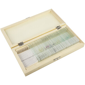
Grundkomponenterna i systemet är de fyra mikroskopiska preparat-skoleserierna A, B, C och D. Serierna är systematiskt ordnade och sammansatta så att de bygger på varandra och utvidgar ämnesområdet för den föregående. Varje enskilt preparat är noggrant utvalt och har granskats med avseende på sitt pedagogiska värde. Vid valet av preparat har de som är typiska för den motsvarande djur- eller växtgruppen getts företräde. Materialet är rikligt tilltaget så att läraren kan välja och variera. LIEDER-mikropreparat tillverkas i våra laboratorier under vetenskaplig ledning. De är resultatet av årtionden av erfarenhet inom alla områden av prepareringsteknik. Mikrotomskärningarna utförs av erfarna specialister, skärteknik och skärtjocklek anpassas efter objekten. Av det stora antalet färgningsmetoder som är vanliga inom mikroskopi väljer vi sådana som ger en tydlig och kontrastrik återgivning av de önskade strukturerna med bästa hållbarhet. Oftast handlar det om komplicerade flerfärgningar. LIEDER-mikroskoppreparat levereras på fint slipade objektglas i formatet 26 x 76 mm. Varje mikroskoppreparat är unikt. Vi vill därför påpeka att levererade preparat kan avvika från bilderna i denna katalog på grund av naturliga variationer i utgångsmaterialet och de preparations- och färgningsmetoder som använts.
Antalet tillgängliga preparatserier, eller åtminstone delar av dessa, bör ungefär motsvara antalet mikroskop så att flera elever samtidigt kan undersöka samma preparat. Därför kan alla mikroskopiska preparat från serierna också beställas enskilt, så att viktiga preparat kan anskaffas i klassstorlek.
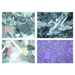
Mognadsdelningar i pollenmodercellerna hos liljan (Lilium)
Serie om fortplantning och ärftlighet
12 preparat.
1. Leptotän. Spiremstadium av kromosomerna. Alla celler med diploida kromosomer.
2. Zygoten. Början av parning av homologa kromosomer
3. Pachytän. Färdiga kromatidpar (Gemini)
4. Diploten. Bildning av chiasmata (korsning), varvid genutbyte och ny kombination av arvsmassan sker
5. Diakines. Spiralformning och därmed förkortning av kromatidentetraderna, slutet av profasstadiet
6. Metafas och anafas av den första (heterotypiska) mognadsdelningen, bildning av kärnspindeln. Det bildas två haploida kromosomuppsättningar
7. Telofas av den första och profasen av den andra (homöotypiska) mognadsdelningen
8. Metafas och anafas av den andra delningen (mitos). Fyra haploida kärnor bildas
9. Pollentetraden efter den andra delningen. Bildning av cellväggar mellan dotterkärnorna
10. Enkärniga (haploida) mikrosporer efter separering av tetradecellerna
11. Tvåkärniga (färdiga) pollenkorn efter den tredje delningen med generativ och vegetativ kärna
12. Mogna pollenkorn, totalt. Ytstruktur
I leveransen och i priset ingår en eller två plastbehållare för 25 preparat vardera. Andra förvaringslådor kan beställas separat.