Tyvärr har den här beskrivningen ännu inte översatts till svenska, så du hittar en engelsk artikelbeskrivning här.
Stains
• Cell staining is a technique that can be used to better visualize cells and cell components under a microscope
• By using different stains, one can preferentially stain certain cell components, such as a nucleus or a cell wall, or the entire cell
• Most stains can be used on fixed, or non-living cells, while only some can be used on living cells; some stains can be used on either living or non-living cells
• The most basic reason that cells are stained is to enhance visualization of the cell or certain cellular components under a microscope
• Cells may also be stained to highlight metabolic processes or to differentiate between live and dead cells in a sample
• Cells may also be enumerated by staining cells to determine biomass in an environment of interest
• There are several types of staining media, each can be used for a different purpose
• Commonly used stains and how they work are listed below
• All these stains may be used on fixed, or non-living, cells and those that can be used on living cells are noted
• After staining cells and preparing slides, they may be stored in the dark and possibly refrigerated to preserve the stained slide
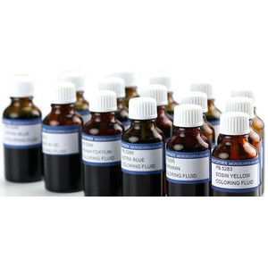
• Supplied in 25 ml bottles
• Information of the dissolved stain can be found in the safety data sheets
• Safety data sheets include information about the properties of the substance (or mixture), its hazards and instructions for handling, disposal and transport and also first-aid, fire-fighting and exposure control measures
Why Stain Cells
The most basic reason that cells are stained is to enhance visualization of the cell or certain cellular components under a microscope. Cells may also be stained to highlight metabolic processes or to differentiate between live and dead cells in a sample. Cells may also be enumerated by staining cells to determine biomass in an environment of interest
What Are Some Common Stains?
There are several types of staining media, each can be used for a different purpose. Commonly used stains and how they work are listed below. All these stains may be used on fixed, or non-living, cells and those that can be used on living cells are noted
• Analin Blue - To be used as third colour for Azo pigmentation (PB.5300)
• Astra Blue - Stain for vegetal cells.To be used in combination with safranin (PB.5283)
• Azo Carmine-G - colors glycogen, biological stains for animal tissues. Also suitable for bacteria pigmentation, red (PB.5280)
• Eosin - a counterstain to haematoxylin, this stain colors red blood cells, cytoplasmic material, cell membranes, and extracellular structures pink or red. Stain for general overall-view colouring, yellow (PB.5283)
• Fuchsin - This stain is used to stain collagen, smooth muscle, bacilli in tissue or mitochondria (PB.5303)
• Hematoxylin - a general purpose nuclear stain that, with a mordant, stains nuclei blue-violet or brown (PB.5286)
• Orange-G - Stain for most elementary structures of animal tissues (PB.5292)
• Methylene blue - stains animal cells to make nuclei more visible. Biological and bacteriological stain (PB.5297)
• Safranin - a nuclear stain used as a counterstain or to color collagen yellow. A general stain for showing nuclei and cellulose walls. To be used in combination with Astra Blue (PB.5295)
After staining cells and preparing slides, they may be stored in the dark and possibly refrigerated to preserve the stained slide, and then observed with a microscope


